Sabtu, 21 Juni 2014

Shigella sp

Diposting oleh Healthy Inside Fesh Outside di 08.39 0 komentar


Shigella
Photomicrograph of Shigella sp. in a stool specimen
Scientific classification
Kingdom:Bacteria
Phylum:Proteobacteria
Class:Gammaproteobacteria
Order:Enterobacteriales
Family:Enterobacteriaceae
Genus:Shigella
Castellani & Chalmers 1919
Species


This article is about the genus. For the disease, see shigellosis
Shigella is a genus of Gram-negativenonspore forming, non-motile, rod-shaped bacteria closely related to Escherichia coli and Salmonella. The causative agent of human shigellosisShigella causes disease in primates, but not in other mammals. It is only naturally found in humans and apes. During infection, it typically causes dysentery. The genus is named after Kiyoshi Shiga, who first discovered it in 1898.
Phylogenetic studies indicate that Shigella is more appropriately treated as subgenus of Escherichia, and that certain strains generally considered E. coli – such as E. coli O157:H7 – are better placed in Shigella (see Escherichia coli#Diversity for details).


Shigella
 species are classified by four serogroups:[edit]
Classification

Groups AC are physiologically similar; S. sonnei (group D) can be differentiated on the basis of biochemical metabolism assays.[4] Three Shigella groups are the major disease-causing species: S. flexneri is the most frequently isolated species worldwide, and accounts for 60% of cases in the developing world; S. sonnei causes 77% of cases in the developed world, compared to only 15% of cases in the developing world; and S. dysenteriae is usually the cause of epidemics of dysentery, particularly in confined populations such as refugee camps.

[edit]Pathogenesis

Shigella infection is typically via ingestion (fecal–oral contamination); depending on age and condition of the host, less than 100 bacterial cells can be enough to cause an infection.  Shigella causes dysentery that results in the destruction of the epithelial cells of the intestinal mucosa in the cecum and rectum. Some strains produce enterotoxin and shiga toxin, similar to the verotoxin of E. coli O157:H7and other verotoxin-producing Escherichia coli. Both shiga toxin and verotoxin are associated with causing hemolytic uremic syndrome. As noted above, these supposed E. coli strains are at least in part actually more closely related to Shigella than to the "typical" E. coli.
Shigella invade the host through the M-cells in the gut epithelia of the large intestine, as they cannot enter directly through the epithelial cells. Using a Type III secretion system acting as a biological syringe, the bacterium injects IpaD protein into cells, triggering bacterial invasion and the subsequent lysis of vacuolar membranes using IpaB and IpaC proteins. It uses a mechanism for its motility by which its IcsA protein triggers actin polymerization in the host cell (via N-WASP recruitment of Arp2/3 complexes) in a "rocket" propulsion fashion for cell-to-cell spread. The most common symptoms are diarrheafevernauseavomiting, stomach cramps and flatulence. The stool may contain blood, mucus, or pus. In rare cases, young children may have seizures. Symptoms can take as long as a week to show up, but most often begin two to four days after ingestion. Symptoms usually last for several days, but can last for weeks. Shigella is implicated as one of the pathogenic causes ofreactive arthritis worldwide.
Each of the Shigella genomes includes a virulence plasmid that encodes conserved primary virulence determinants. The Shigella chromosomes share most of their genes with those of E. coli K12 strain MG1655.

[edit]Diagnosis

Shigella species are negative for motility and are not lactose fermenters. (However, S. sonnei can ferment lactose). They typically do not produce gas from carbohydrates (with the exception of certain strains of S. flexneri) and tend to be overall biochemically inert. Shigella should also be urea hydrolysis negative . When inoculated to a triple sugar iron (TSI) slant, they react as follows: K/A, gas -, H2S -. Indole reactions are mixed, positive and negative, with the exception of S. sonnei, which is always indole negative. Growth on Hektoen enteric agar will produce bluish-green colonies for Shigella and bluish-green colonies with black centers for Salmonella.

[edit]Treatment

Severe dysentery can be treated with ampicillin, TMP-SMX, or fluoroquinolones, such as ciprofloxacin, and of course rehydration. Medical treatment should only be used in severe cases. Antibiotics are usually avoided in mild cases because some Shigella are resistant to antibiotics, and their use may make the germ even more resistant. Antidiarrheal agents may worsen the sickness, and should be avoided. For Shigella-associated diarrhea, antibiotics shorten the length of infection.

HAEMOGLOBINE

Diposting oleh Healthy Inside Fesh Outside di 08.31 0 komentar


Haemoglobin adalah kompleks protein-pigmen yang mengandung zat besi. Kompleks tersebut berwarna merah dan terdapat didalam eritrosit. Sebuah molekul haemoglobin memiliki empat gugus haeme yang mengandung besi fero dan empat rantai globin. Terdapat beberapa bentuk haaemoglobin : tipe fetal (HbF)  dan dua bentuk utama haemoglobin dewasa (HbA1 dan HbA2). Haemoglobin membawa oksigen, sebagian karbondioksida dan mendapat perubahan pH.
Glycosylated haemoglobin (HbA1) ---> kadar HbA1 menunjukkan kadar gula darah selama periode beberapa bulan dan dapat digunakan untuk menilai derajat pengendalian pada Diabetes mellitus.
Nilai normal Hb untuk laki-laki adalah 13 gr% - 18 gr%, dan untuk wanita adalah 11,5 gr% - 16,5 gr% (Brooker, 2001). 


Haemoglobin adalah Sebuah substansi didalam sel darah merah (erithrocyte) dan tanggung jawab masing-masing warna, terdiri dari pigmen haeme (zat besi - berisi porphyrin) terkait dengan protein globin. Haemoglobin memiliki sifat unik dapat menyatu dengan oksigen dan merupakan pengangkut oksigen ke seluruh tubuh. Haemoglobin membawa oksigen dalam aliran darah melewati paru-paru dan bersama dengan darah sampai ke jaringan tubuh. Darah biasanya mengandung 12-18 g / dl dari hemoglobin.


Myohaemoglobin : zat besi - yang mengandung protein, menyerupai hemoglobin, ditemukan dalam sel otot. Seperti hemoglobin yang berisi kumpulan haeme. Ikatan yang mengandung oksigen, bertindak sebagai reservoir oksigen di dalam serabut otot .
Oxyhaemoglobin : substansi darah merah dibentuk bila pigmen hemoglobin dalam sel darah merah menyatu kembali dengan oksigen. Oxyhaemoglobin adalah bentuk oksigen yang diangkut dari paru-paru ke sel-sel, di mana oksigen dilepaskan.
Methahaemoglobin : substansi yang dibentuk apabila atom besi dari pigmen hemoglobin darah telah mengoksidasi dari ferrous ke bentuk ferric (bandingkan oxyhaemoglobin). Methahaemoglobin yang tidak dapat mengikat oksigen molekular dan karenanya tidak dapat mentransportasi oksigen ke seluruh tubuh. Keberadaan methahaemoglobin dalam darah (methahaemoglobinaemia) mungkin akibat menelan zat oksid dari narkoba atau dari warisan keabnormalan dari molekul hemoglobin. Gejala - gejala termasuk kelelahan, sakit kepala, pusing dan cyanosis (oxford electric medical dictionary).

Referensi :
Brooker, Christine. 2001. Kamus Saku Keperawatan.EGC  : Jakarta.
oxford electric medical dictionary (Indonesian Translete by Patriani)
English Version

Haemoglobin is the pigment-protein complex that contains iron. Complex is red and there is the eritrosit. A haemoglobin molecule has four haeme group which contains iron and fero four globin chains. There are several forms of haaemoglobin: type fetal (HbF) and the two main forms of adult haemoglobin (HbA1 and HbA2). Haemoglobin takes oxygen, some carbon dioxide and the pH changes. Glycosylated haemoglobin (HbA1) ---> HbA1 level indicates the blood sugar over a period of several months and can be used to assess the degree of control on Diabetes mellitus. Hb normal value for men is 13% gr - 18 gr%, and for women is gr 11.5% - 16.5 gr% (Brooker, 2001).

Haemoglobin is a substance contained within the red blood cell (erithrocyte) and responsible for their colour, composed of the pigment haem (an iron - containing porphyrin) linked to the protein globin. Haemoglobin has the unique property of combining reversibly with oxygen and is the medium by which oxygen is transported within the body. it take up oxygen as blood passes through the lungs and releases it as blood passes through the tissues. Blood normally contains 12-18 g/dl of haemoglobin.

Myohaemoglobin : an iron – containing protein, resembling haemoglobin, found in muscle cell. Like haemoglobin it contains a haem group. Which binds reversibly with oxygen, and so acts as an oxygen reservoir within the muscle fibres.

Oxyhaemoglobin : the bright – red substance formed when the pigment haemoglobin in red blood cell combines reversibly with oxygen. Oxyhaemoglobin is the form in which oxygen is transported from the lungs to the tissues, where the oxygen is released.

Methahaemoglobin : a substance formed when the iron atoms of the blood pigment haemoglobin have been oxidized from the ferrous to the ferric form (compare oxyhaemoglobin). The methahaemoglobin cannot bind molecular oxygen and therefore it cannot transport oxygen round the body. The presence of methahaemoglobin in the blood (methahaemoglobinaemia) may result from the ingestion of oxidizing drugs or from an inherited abnormality of the haemoglobin molecule. Symtomps include fatigue, headache, dizziness and cyanosis (oxford electric medical dictionary).


Japanese Version

モグロビンは鉄を含んでいる顔料蛋白質の複合体である。 複合体は赤く、eritrositがある。 ヘモグロビンの分子に鉄およびferoを4つのglobinの鎖含んでいる4つのhaemeのグループがある。 haaemoglobinの複数の形態がある: 胎児タイプ(HbF)および大人のヘモグロビンの2つの主要な形態(HbA1およびHbA2)。 ヘモグロビンは酸素、二酸化炭素およびpH変更を取る。 Glycosylatedヘモグロビン(HbA1) ---> HbA1レベルは数月一定期間に渡って血糖を示し、糖尿病mellitusの制御のある程度を査定するのに使用することができる。 人のためのHbの正常な価値は13% gr -女性のための18 gr%、gr 11.5%はあり- 16.5 gr%である。(Brooker, 2001).

ヘモグロビンです 物質は、赤血球( erithrocyte )とその色は、色素裾(鉄-ポルフィリンを含む)は、タンパク質のグロビンにリンクに含まれる構成を担当。可逆ヘモグロビンの酸素と結合しているとのユニークな特性は、酸素が体内の酸素運搬されている媒体です。血液が肺を通過すると、血液の組織を通過するとしても酸素をリリースする。血液は通常12から18グラム/ヘモグロビンのライブラリが含まれて.

Myohaemoglobin: 鉄-ヘモグロビンに類似している蛋白質を含んでいて筋肉細胞で見つけた。 ヘモグロビンのようにそれはhaemグループを含んでいる。 、および従ってリバーシブルに結合するかどれが酸素と機能する筋繊維内の酸素の貯蔵所として。

Oxyhaemoglobin: 明るい – 赤物質、色素ヘモグロビン赤い血のセルに組み合わせた可逆的酸素とを形成します。 Oxyhaemoglobin はで酸素に運ばれる、肺から、組織、酸素が解放されます。

Methahaemoglobin: 物質形成を ferric フォーム (比較 oxyhaemoglobin) に、鉄原子血液色素ヘモグロビンが、鉄から酸化されている場合。 methahaemoglobin、酸素の分子をバインドできませんおよびラウンド、身体の酸素をトランスポートとことはできませんしたがってします。 ヘモグロビン分子の遺伝的異常から、または、経口摂取の薬物を酸化剤から methahaemoglobin (methahaemoglobinaemia)、血液中の存在があります。 Symtomps には、疲労、頭痛、めまい、および cyanosis が含まれます。(oxford electric medical dictionary) 

参照 
Brooker, Christine。 2001。 Kamus Saku Keperawatan. EGC : Jakaruta. 
oxford 電気医療辞書 (日本語翻訳 Translete.Net によって)

HIJAB

Diposting oleh Healthy Inside Fesh Outside di 08.13 0 komentar


Merasa enggak pede saat mix and matchhijab dengan outfit favorit? Saat mengenakan hijab, terutama buat pemula enggak perlu khawatir menggabungkan berbagai gaya supaya penampilan kita tambah oke. Berbusana hijab bukan berarti enggak bisa mengikuti tren. Selama kita pintar mix and match hijab dengan outfit favorit, kita bisa tampil stylish dengan cantik. Simak yuk panduan mix and match hijab dengan outfit favorit!

Polos
Outfit polos paling mudah kita mix and match saat menggunakan hijab. Kuncinya, memilih warna dan material yang tepat. Kita bisa mengkombinasikan oufit polos dengan teknik layering atau pemilihan gaya simple agar tetap nyaman walaupun cuaca sedang panas. Pilih warna netral agar mudah kitamatch dengan outfit lain. Atau gunakan satu warna mencolok diantara warna netral agar penampilan enggak tabrak warna dan terlihat selaras.

Hijab Motif
Yang aman saat menggunakan hijab motif adalah pakai atasan polos. Mengkombinasikan hijab motif enggak terlalu sulit selama kita mengikuti aturan yang benar. Untuk bagian celana atau rok boleh saja menggunakan pakaian bermotif. Asal warnanya senada dengan warna hijab yang kita pakai. Kombinasikan atasan kita dengan material unik supaya tambah menarik. Misalnya kemeja denim, bahan sifon atau kaos dengan detail lucu.

Atasan Motif
Ketika memilih atasan motif, artinya hijab kita sebaiknya berwarna polos. Warna polos akan menyeimbangkan dan memperlihatkan motif pada atasan kita semakin kuat. Saat menyesuaikan warna hijab, enggak harus senada dengan atasan atau bawahan. Kita bisa mengkreasikannya dengan warna cerah lain asal matching dengan warna yang kita pakai. Misalnya hijab oranye dengan atasan biru dan celana jeans.

Printed Hijab on Printed Top
Kalau kita mau eksperimen gaya dengan atasan dan hijab yang bermotif, caranya padukan alternative outfit kita dengan pakaian polos. Kita bisa memadukan atasan motif pakai cardigan atau blazer polos. Bisa juga menggunakan layering dengan menambahkan inner atau outer polos saat kita memadukannya dengan hijab colorful.

Sabtu, 14 Juni 2014

FISIOLOGI GINJAL

Diposting oleh Healthy Inside Fesh Outside di 07.37 0 komentar
A.    Letak
Manusia memiliki sepasang ginjal yang terletak di belakang perut atau abdomen. Ginjal ini terletak di kanan dan kiri tulang belakang, di bawah hati dan limpa. Di bagian atas (superior) ginjal terdapat kelenjar adrenal (juga disebut kelenjar suprarenal).
            Ginjal adalah sepasang organ saluran kemih yang terletak di rongga retroperitoneal bagian atas. Bentuknya menyerupai kacang dengan sisi cekungnya menghadap ke medial. Kedua ginjal terletak di sekitar vertebra T12 hingga L3. Ginjal kanan biasanya terletak sedikit di bawah ginjal kiri untuk memberi tempat untuk hati.
Sebagian dari bagian atas ginjal terlindungi oleh iga ke sebelas dan duabelas. Kedua ginjal dibungkus oleh dua lapisan lemak (lemak perirenal dan lemak pararenal) yang membantu meredam goncangan.
B.     Struktur detail
Berat dan besar ginjal bervariasi; hal ini tergantung jenis kelamin, umur, serta ada tidaknya ginjal pada sisi lain.Pada orang dewasa, rata-rata ginjal memiliki ukuran panjang sekitar 11,5 cm, lebar sekitar 6 cm dan ketebalan 3,5 cm dengan berat sekitar 120-170 gram atau kurang lebih 0,4% dari berat badan. Ginjal memiliki bentuk seperti kacang dengan lekukan yang menghadap ke dalam. Di tiap ginjal terdapat bukaan yang disebut hilus yang menghubungkan arteri renal, vena renal, dan ureter.
C.    Vaskularisasi
Aliran darah ginjal berasal dari arteri renalis yang merupakan cabang langsung dari aorta abdominalis, sedangkan yang mengalirkan darah balik adalah vena renalis yang merupakan cabang vena cava inferior. Sistem arteri ginjal adalah tidak ada anastomosis ke cabang arteri lain
D.    Bagian-bagian Ginjal
Bagian paling luar dari ginjal disebut korteks, bagian lebih dalam lagi disebut medulla. Bagian paling dalam disebut pelvis. Pada bagian medulla ginjal manusia dapat pula dilihat adanya piramida yang merupakan bukaan saluran pengumpul. Ginjal dibungkus oleh jaringan fibros tipis dan mengkilap yang disebut kapsula fibrosa ginjal dan diluar kapsul ini terdapat jaringan lemak perirenal. Di sebelah atas ginjal terdapat kelenjar adrenal. Ginjal dan kelenjar adrenal dibungkus oleh fasia gerota. Unit fungsional dasar dari ginjal adalah nefron yang dapat berjumlah lebih dari satu juta buah dalam satu ginjal normal manusia dewasa. Nefron berfungsi sebagai regulator air dan zat terlarut (terutama elektrolit) dalam tubuh dengan cara menyaring darah, kemudian mereabsorpsi cairan dan molekul yang masih diperlukan tubuh. Molekul dan sisa cairan lainnya akan dibuang. Reabsorpsi dan pembuangan dilakukan menggunakan mekanisme pertukaran lawan arus dan kotranspor. Hasil akhir yang kemudian diekskresikan disebut urine. Sebuah nefron terdiri dari sebuah komponen penyaring yang disebut korpuskula (atau badan Malphigi) yang dilanjutkan oleh saluran-saluran (tubulus). Setiap korpuskula mengandung gulungan kapiler darah yang disebut glomerulus yang berada dalam kapsula Bowman. Setiap glomerulus mendapat aliran darah dari arteri aferen. Dinding kapiler dari glomerulus memiliki pori-pori untuk filtrasi atau penyaringan. Darah dapat disaring melalui dinding epitelium tipis yang berpori dari glomerulus dan kapsula Bowman karena adanya tekanan dari darah yang mendorong plasma darah. Filtrat yang dihasilkan akan masuk ke dalan tubulus ginjal. Darah yang telah tersaring akan meninggalkan ginjal lewat arteri eferen.
 Di antara darah dalam glomerulus dan ruangan berisi cairan dalam kapsula Bowman terdapat tiga lapisan:
1.      kapiler selapis sel endotelium pada glomerulus
2.      lapisan kaya protein sebagai membran dasar
3.      selapis sel epitel melapisi dinding kapsula Bowman (podosit)

Dengan bantuan tekanan, cairan dalan darah didorong keluar dari glomerulus, melewati ketiga lapisan tersebut dan masuk ke dalam ruangan dalam kapsula Bowman dalam bentuk filtrat glomerular. Filtrat plasma darah tidak mengandung sel darah atau pun molekul protein yang besar. Protein dalam bentuk molekul kecil dapat ditemukan dalam filtrat ini. Darah manusia melewati ginjal sebanyak 350 kali setiap hari dengan laju 1,2 liter per menit, menghasilkan 125 cc filtrat glomerular per menitnya. Laju penyaringan glomerular ini digunakan untuk tes diagnosa fungsi ginjal.

Jumat, 13 Juni 2014

How to apply a simple Make up

Diposting oleh Healthy Inside Fesh Outside di 07.40 1 komentar
STEP 1


Use good face cleaners. Wash your face. Washing your face will make your skin look perfectly clean. This cleans your face well and gets rid of dead skin, blackheads, oil, and dirt. Make sure you don't clean your face with aggressive cleaners or soaps. Use a cleanser that is complementary for your skin, for advice go to your dermatologist or ask for advice in a store where they sell beauty supplies.

STEP 2

Do Your Makeup Flawlessly Step 2.jpg

Find a good moisturizer. Apply a moisturizer. Pick a moisturizer that fits with your skin type:
  • Dry/sensitive skin: choose a thicker cream/ointment that locks moisture in.
  • Normal skin: choose cream moisturizers that are hydrating.
  • Oily/young skin: choose a water based lotion.
STEP 3

Do Your Makeup Flawlessly Step 3.jpg

Use a good primer, any brand will do. Add a foundation primer. Foundation primers are used to set make up (foundation) so that it lasts longer.You can find several in drug stores or specialized beauty stores.

STEP 4

Do Your Makeup Flawlessly Step 4.jpg

Choose your foundation. If you already have flawless skin (no freckles, blemishes, discoloration, or uneven tone) you should use a tinted moisturizer. This will add a bit of color without looking cake-like. If you want to cover a bit more, use a foundation.This is one product that you should splurge on. To get the right shade apply some near your cheek in natural daylight and make sure to test a bunch of them to find the right one for you. Ask one of the professionals in the store to help you find the right color.
  • Apply the foundation. To get your skin dewy and radiant use an illuminator. Just mix a pea-sized amount of illuminator with your foundation and you'll get glowing skin. Apply the foundation with a foundation brush if you have one or use your fingers. Some people like using brushes, but you may prefer fingers because it's easier, just remember to wash your hands before. Put a bit of foundation on the back of your hand, dab a bit of foundation onto your fingers and smooth it onto your face. Make sure to get your jawline, ears and around your eyes. Remember: foundation is meant to even out your skin tone. Don't use it to add color or completely cover blemishes.

STEP 5

Do Your Makeup Flawlessly Step 5.jpg

Buy a good concealer. Search for a concealer close to your skin tone, making sure you don't choose one that is too dark or too light. If you can't tell, ask for advice to ensure you get the right color.
  • Apply the concealer on any zits or redness on your skin , use a small brush to stipple it on and dab it with your fingers to blend it in.
  • Conceal any dark circles under your eye by applying the concealer in a triangle shape. Blend the outer corners of the triangle with your fingers. Don't forget to conceal the inner corners of your eyes as they are generally darker than the rest of your eyes.
STEP 6

Do Your Makeup Flawlessly Step 6.jpg

Set the concealer with setting powder. Choose a loose (mineral) powder and dab it on the concealed parts of your face with a stippling brush.

STEP 7

Do Your Makeup Flawlessly Step 7.jpg

Apply a bronzer and/or blush. Choose a bronzer that isn't too dark for your skin color otherwise you will end up looking like a clown. Use an angled, hard brush or a stippling brush to apply the bronzer, work in a cross on your face (forehead, nose and cheeks). To make your face look slimmer apply the bronzer right under the apples on your cheeks, to find the apples smile and apply the bronzer in the hollow of your cheeks.
  • Find a blush that suits your skin tone and apply it on the apples of your cheeks.
  • To prevent your face becoming shiny you can use oil blotting sheets. You can find them in any drug store or store where they sell beauty products. They're cheap, reduce shine, and don't disturb makeup. Bring these along with you.

STEP 8
Do Your Makeup Flawlessly Step 8.jpg


Apply eye makeup. To make your eyeshadow last, apply an eye shadow primer first. For natural eye looks choose a natural color.
  • Curl your lashes. Curl your eyes for about 15 seconds on each side.
  • Use an eyelash extender. The fibers from this connect and make lashes much longer.
  • Apply a waterproof mascara. Wait for each coat to dry before applying another to avoid clumps.
  • Apply eyeliner. Liquid or pencil, top or bottom, eyeliner can be applied in many ways and looks good in different ways depending on the eye shape, color, and size. You'll really just have to experiment with this.
    • Color your brows if they aren't even with a eyebrow powder, choose a color that is close to your eyebrow color. Apply a brow gel to keep your eyebrows in place.
STEP 10
Do Your Makeup Flawlessly Step 9.jpg

Moisturize your lips by applying chapstick. Use a lip primer to fill up any creases and apply a lip liner, followed by applying a lipstick in the same color as the lip liner and finish with a gloss if necessarily.

STEP 10 Do Your Makeup Flawlessly Intro.jpg


Fasciola Hepatica - Liver Fluke

Diposting oleh Healthy Inside Fesh Outside di 07.06 0 komentar

Fasciola Hepatica - Liver Fluke

Fasciola hepatica is a parasitic fluke that lives in the liver. In addition to humans it infects cows and sheep. It is known as the common liver fluke and causes a disease called fascioliasis.
The life cycle of Fasciola hepatica starts when a female lays eggs in the liver of an infected human. Immature eggs are discharged in the biliary ducts and taken out in the feces. If landed in water, the eggs become embryonated and develop larvae called miracidia. A miracidium invades an aquatic snail and develops into cercaria, a larva that is capable of swimming with its large tail. The cercaria exits and finds aquatic vegetation where it forms a cyst called metacercaria. A human eats the raw freshwater plant containing the cyst. The metacercaria excysts in the first part of the small intestine, duodenum. It then penetrates the intestinal wall and gets into the peritoneal cavity. It finds the liver and starts eating liver cells. This happens only a few days after the initial contact with the parasite. Usually the larva spends a few weeks just browsing and eating the liver. Then it relocates to the bile duct where it begins its final stage and becomes an adult. It takes about three months for the metacercaria to develop into an adult. Adults are about 3 cm long and 1 cm wide. Adult females can produce up to 25000 eggs per day.
In the chronic phase of fascioliasis adults in the large biliary ducts cause liver inflammation and obstruction of the biliary fluid. During the migration of the larvae (this acute phase of the disease lasts many weeks) symptoms include:
  • diarrhea
  • eosinophilia (high number of white blood cells)
  • fever
  • nausea
  • stomach ache
  • vomiting.
Fasciola hepatica is found in areas where cattle and sheep are raised.
Fasciola hepatica is identified from eggs in a stool sample. The eggs are very similar to those of Fasciolopsis buski. Early stage of the infection can be diagnosed from a blood sample, if antibodies are found. Fascioliasis is treated with triclabendazole drug.

             Fasciola hepatica life cycle

Fasciola hepatica body parts     Fasciola hepatica

Fasciola hepatica life cycle

Diposting oleh Healthy Inside Fesh Outside di 07.03 0 komentar

Causal Agent:

Fascioliasis is caused by Fasciola hepatica and less often by F. gigantica, which are flat worms classified as liver flukes (trematodes). Some human cases have been caused by hybrid species. Additional Fasciola species have been found in animals.

Life Cycle:

As shown below, Fasciola parasites develop into adult flukes in the bile ducts of infected mammals, which pass immature Fasciola eggs in their feces. The next part of the life cycle occurs in freshwater. After several weeks, the eggs hatch, producing a parasite form known as the miracidium, which then infects a snail host. Under optimal conditions, the development process in the snail may be completed in 5 to 7 weeks; cercariae are then shed in the water around the snail. The cercariae lose their tails when they encyst as metacercariae (infective larvae) on water plants. In contrast to cercariae, metacercariae have a hard outer cyst wall and can survive for prolonged periods in wet environments.
Life cycle of Fasciola

Immature Fasciola eggs are discharged in the biliary ducts and in the stool The number 1
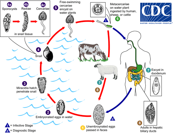
. Eggs become embryonated in water The number 2, eggs release miracidia The number 3, which invade a suitable snail intermediate host The number 4, including the genera Galba, Fossaria and Pseudosuccinea. In the snail the parasites undergo several developmental stages (sporocysts The number 4a, rediae The number 4b, and cercariae The number 4c). Thecercariae are released from the snail The number 5 and encyst as metacercariae on aquatic vegetation or other surfaces. Mammals acquire the infection by eating vegetation containing metacercariae. Humans can become infected by ingesting metacercariae-containing freshwater plants, especially watercress The number 6. After ingestion, the metacercariae excyst in the duodenum The number 7 and migrate through the intestinal wall, the peritoneal cavity, and the liver parenchyma into the biliary ducts, where they develop into adult flukes The number 8.
In humans, maturation from metacercariae into adult flukes takes approximately 3 to 4 months. The adult flukes (Fasciola hepatica: up to 30 mm by 13 mm; F. gigantica: up to 75 mm) reside in the large biliary ducts of the mammalian host. Fasciola hepatica infect various animal species, mostly herbivores (plant-eating animals).

adapted : http://www.cdc.gov/parasites/fasciola/biology.html
 

KESEHATAN DAN KECANTIKAN Copyright © 2012 Design by Antonia Sundrani Vinte e poucos